Bone Augmentation Upper and Lower Jaw

The content provided here is intended solely for neutral information and general educational purposes. It does not constitute a recommendation or endorsement of the diagnostic methods, treatments, or medications described. This text does not replace professional advice from a dentist, physician, or pharmacist and must not be used as a basis for self-diagnosis or for initiating, modifying, or discontinuing any treatment. If you experience dental, oral, or jaw-related issues, or have any other health concerns, always consult a qualified dentist or healthcare professional.
If the bone height and width are insufficient for a dental implant to be placed, bone augmentation may need to be performed in the upper and lower jaw.
There are many different techniques, methods, and materials to restore the missing volume. It is essential to ensure a healthy and stable solution.
A special type of bone augmentation in the upper jaw is the maxillary sinus lift.
In the following, we would like to elaborate on the various possibilities and reasons for bone augmentation.
What is bone augmentation?
Bone augmentation (in our dental context) refers to building up the jawbone. Jaw bone augmentation procedures may be essential before or during implant treatment to ensure the long-term success, safety, and stability of the implants and to create appealing aesthetics, or it can also be used to support the periodontium. Various materials and methods are used for this purpose.
Reasons for bone augmentation
If bone loss has occurred prior to treatment and the planned therapy requires a sufficient foundation, a bone augmentation is necessary. The causes of bone loss are diverse.
The human body follows the principle of “use it or lose it.” If bone is no longer loaded by chewing forces (by a complex interplay of pressure and tension mechanisms), it begins to shrink. These situations may necessitate bone augmentation:
Periodontitis / Periodontal Disease
When bacterial inflammation (periodontitis/periodontal disease) has weakened the periodontal apparatus over a long period, it can lead not only to gum recession, but also to bone defects. When affected teeth are then removed, the bone level is already too low from the beginning on. Traumatic extractions using excessive force increase the extent of losses. Before implant treatment, these defects must be compensated. Depending on the situation, a prosthodontic restoration (such as a ceramic bridge) may also require bone augmentation if there are aesthetic limitations due to the loss.
Tooth extraction without socket preservation
After tooth removal without accompanying bone-preserving measures (socket preservation), it may come to a significant loss of bone volume within the first 6-12 months. Depending on the initial situation and the characteristics of the bone, the jawbone may lose up to 50% of its original width, which can complicate subsequent implant treatment.
Tooth removal without implant placement
If a gap remains untreated for a long time, bone loss can occur over time. The jawbone needs regular activating chewing forces to maintain its structure and density. Without this stimulation by tooth roots or implants, the bone may recede.
Long-term wearing of a denture
Removable dentures (partial or complete dentures) can load additional stress on the underlying jawbone. The high pressure on the jaw can lead to an accelerated bone loss, especially if the denture is not optimally adjusted, pressure points exist, or if it is worn for many years.
Bone augmentation for maintaining natural teeth
Bone augmentation is not only relevant for dental implant treatments, but can also be performed on natural teeth. The goal is to gain bone substance around healthy natural teeth.
Regenerative therapy for periodontitis
In cases of advanced periodontitis, special regenerative procedures can be used to rebuild lost bone around natural teeth. By using bone substitute materials, possibly enamel matrix proteins, in combination with special membranes (Guided Tissue Regeneration, GTR), the bone volume can be specifically built up.
Bone augmentation after dental trauma
After accidents with injuries to the jawbone, bone augmentation may be necessary to preserve the natural teeth or to enable later prosthodontic treatment.
In preparation for orthodontic treatments
Sometimes bone augmentation is required before orthodontic procedures can be performed in order to create a sufficient basis for tooth movements. If this is omitted, exposed root surfaces are possible, which limit the long-term prognosis of the teeth.
Bone augmentation for implant placement
For the successful insertion and long-term stability of dental implants, an adequate bone supply is essential, as it forms the basis of the restoration. Various methods are available depending on the situation and extent of the bone defect.
Sinus floor elevation
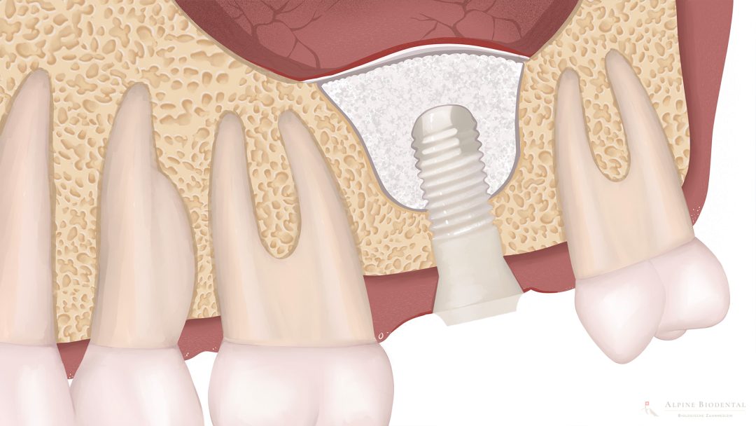
The sinus lift is a well known method for bone augmentation in the upper jaw posterior region. The floor of the maxillary sinus (sinus maxillaris) is elevated, and the created space is filled with bone substitute material, autogenous bone, or directly with the dental implant.
Two techniques are distinguished:
- External sinus lift - direct sinus floor elevation: For larger defects, the maxillary sinus is accessed through a lateral window. This method enables larger bone augmentations.
- Internal sinus lift - indirect sinus floor elevation: With residual bone height of 5 - 8 mm, the sinus floor can be lifted through the implant access without a separate access window. This technique is minimally invasive.
Bone block augmentation
For significant bone defects, a bone block graft may be the best solution. A block of autogenous bone (usually from the jaw, chin, or iliac crest) is removed and firmly screwed in the defect area. After its integration, implant placement can be performed at a later time. This method offers high biological value and good stability, however, the, blood supply may be limited.
Bone splitting / Bone spreading
With sufficient bone height but insufficient bone width, the existing bone can be carefully expanded:
- Bone Splitting: The alveolar ridge is split longitudinally and spread to create space for implants.
- Bone Spreading: The bone is gently and gradually stretched and compacted, thus stabilized.
Both techniques expand the narrow alveolar ridge and create additional space for implants without having to remove bone from other sites.
Socket preservation
Socket preservation is a preventive measure immediately after tooth removal. The empty socket is filled with bone substitute material and usually covered with a membrane. This technique minimizes bone loss and preserves bone volume for later implant restoration.
Distraction osteogenesis
This sophisticated special procedure uses the body’s own bone formation: The bone is surgically cut and then slowly stretched over a period of several weeks by a special device. New bone builds in the resulting gap. This technique is used for larger defects.
Materials for bone augmentation
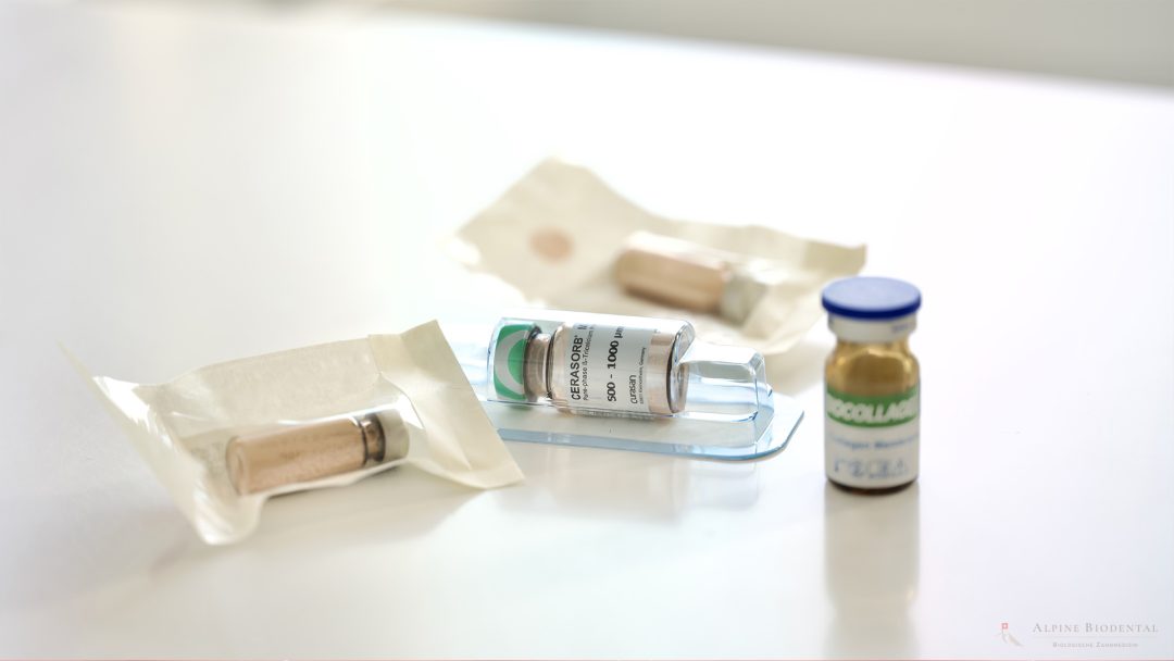
Various materials are available for bone augmentation, which are selected depending on the situation, desired outcome, and personal factors. When choosing, individual circumstances should be considered. Properties such as biology and compatibility, but also bone stability, are of the highest priority.
Autogenous bone - the patient’s own bone
Autogenous bone (the patient’s own bone) is considered to be the “gold standard” due to its biological properties:
- Contains living bone cells and growth factors
- Offers optimal compatibility (no rejection)
- Actively promotes new bone formation
- Serves as a guide for new bone growth
Donor sites are usually the jaw, the chin area, or for larger amounts, the iliac crest. The bone block is gently and precisely removed using piezosurgery and then placed at the correct location. It improves the bone supply in horizontal and vertical directions.
The disadvantage lies in the additional procedure for removal and potential discomfort at the donor site.
Bone chips can also be used as autogenous bone. The bone chips are preferably collected in a sterile bone collector (e.g., Safescraper) and are placed in the bone defect without contamination. Often, the bone chips can be collected at the site of the operation without having to create an additional wound.
Allogeneic bone substitute - Donor bone
Allogeneic bone substitute comes from human donors and is extensively cleaned and sterilized:
- Available in various forms (blocks, granules, etc.)
- Slow, reliable conversion to the body's own bone
- Unlimited quantities available
- No second procedure needed for removal
- Similar structure to own bone
Due to special preparation processes, the risk of disease transmission or intolerance is low.
Xenogenic bone substitute - Animal origin
These materials usually come from cattle or pigs, with the organic components removed and also undergo elaborate cleaning and sterilization processes:
- Available in various forms (blocks, granules, etc.)
- Slow, reliable conversion to the body's own bone
- Unlimited quantities available
- No second procedure needed for removal
- Particularly long-term stable scaffold structure
The mineral matrix serves as a guiding structure for the body’s own bone, which grows into the material.
Alloplastic bone substitute - Artificial materials
Synthetically produced bone substitute materials usually consist of compounds of calcium and phosphate:
- No biological origin (important for ethical or religious concerns)
- Controlled production in consistent quality
- Good predictability of healing behavior
These materials are available in various compositions that offer different degradation speeds and mechanical properties.
Healing process after bone augmentation
The healing process after bone augmentation is a complex biological process that requires time and patience. Understanding this process helps to develop realistic expectations.
After introducing the augmentation material, the body goes through several phases:
- Inflammatory phase (first days): The body reacts with a controlled inflammatory response that initiates healing processes. Swelling and mild pain are normal.
- Revascularization (from 1-2 weeks and longer): New blood vessels grow into the bone and supply the area with nutrients and oxygen.
- Osseointegration (2-6 months): The body’s own bone cells colonize the material and begin the remodeling process. This process is also known as “creeping substitution,” as the bone substitute material is gradually replaced by the body’s own bone.
- Remodeling (up to 1 year): The new bone functionally adapts to the load and receives its final structure.
The total duration until complete integration depends on various factors:
- Type and extent of bone augmentation
- Shape of the defect
- Material used
- Individual healing of the patient
- Accompanying diseases such as diabetes or osteoporosis
Complications after bone augmentation
As with any surgical procedure, complications can occur during bone augmentation. However, with careful planning, modern technology, and experience of the practitioner, the risk is low.
Possible complications in the upper jaw:
- Opening of the maxillary sinus during sinus lift operations
- Sinusitis (sinus infection)
- Swelling of the mucous membrane, which hinders drainage of the maxillary sinus
- Bleeding into the maxillary sinus
Possible complications in the lower jaw:
- Impairment of the mandibular nerve with temporary or rarely permanent sensory disturbances of the tongue or lower lip
- Possibly increased risk of fracture with extensive procedures
General complications:
- Wound healing disorders or infections
- Subsequent bleeding
- Insufficient integration and loss of the bone augmentation
- Premature degradation of the bone augmentation material
If problems such as persistent pain, severe swelling, bleeding, or fever occur, the treating dentist should be contacted immediately.
Alternatives to bone augmentation
An elaborate bone augmentation is not required in every situation. There are various alternatives that can be considered depending on the individual situation. It is important to discuss the alternatives in detail with the patient to find the individually best solution together.
Short implants without bone augmentation
Modern short implants (6-8 mm length) can be used in many cases even with reduced bone supply:
- Avoidance of elaborate augmentations
- Shorter treatment time
- Lower costs
- Less invasive
It is important to note the distribution of chewing forces, as short implants distribute lateral pressure over a smaller surface than longer implants.
Careful control of prosthetic contact points and the patient’s chewing function is essential to avoid overloading.
Angled implants
By angling the implants, anatomical structures such as the sinus in the upper jaw or the nerve canal in the lower jaw may be bypassed:
- Utilization of the existing bone
- Avoidance of augmentations
- Usually only applicable with multiple implants and special restoration concepts
Treatment with a ceramic bridge
With sufficiently stable neighboring teeth, a fixed cemented bridge can be a good alternative:
- No surgical intervention necessary
- Faster restoration
- Aesthetically pleasing results through modern ceramics
- No waiting time for healing phases
However, (healthy) neighboring teeth must be drilled on, which means an irreversible loss of substance and also causes stronger pressure loading through chewing forces.
Removable dentures
A removable partial or complete denture can provide a functional solution:
- No surgical interventions necessary
- Cost-effective
- Often easy to use even with little bone supply
- Regular adjustments possible
- Expandable solution if more teeth need to be removed in the future
The disadvantage lies in the lower chewing function and possible loss of comfort compared to fixed solutions. Many patients complain about the foreign body sensation and do not feel comfortable with a denture.
Dr. med. dent. Josephine Phillips
Bone augmentation may be necessary before an implantation. The correct material and a careful approach are crucial.

Frequently asked questions about bone augmentation
Patients often have many questions about bone augmentation. Here are answers to the most frequently asked questions.
The waiting time depends on the extent of bone augmentation and the material used:
- For smaller bone augmentations and simultaneous implantation: No additional waiting time
- For medium to larger augmentations: 3-6 months
- For extensive bone block augmentations: 5-6 months
Individual healing ability and anatomy of the respective region also play an important role. The ideal time for placing the dental implant after bone augmentation can be determined through clinical monitoring and, if necessary, radiological examinations.
The nutrient supply of the augmented bone occurs through the ingrowth of blood vessels. This process is known as revascularization and is crucial for success:
Autogenous bone already brings living cells, while bone substitute materials must first be colonized.
A healthy diet, adequate vitamin D supply, and abstaining from nicotine positively support this process.
Experiences with modern bone augmentation procedures are predominantly positive:
- In the upper jaw: Especially sinus lift procedures show very high success rates
- In the lower jaw: Bone augmentation procedures also have good prognoses, but may require longer healing times due to the denser bone structure
- Horizontal bone augmentations heal more reliably than vertical augmentations, depending on the extent
In the upper jaw, the following average healing times are expected:
- Internal sinus lift with simultaneous implantation: 3-4 months
- Extensive external sinus lift: 4-6 months (implantation may only be possible after this healing period)
- Horizontal augmentations: 4-6 months
The upper jawbone is less dense than the lower jaw, which often leads to faster vascularization, but can also mean faster volume loss.
The following guidelines apply to the lower jaw:
- Smaller augmentations: 3-4 months
- Block augmentations: 5-6 months
- Vertical augmentations: 5-6 months
The denser bone structure in the lower jaw can lead to longer healing times but provides a very stable base after successful integration.
The costs vary greatly depending on the extent, technique, and material, so a general statement cannot be made here.
After an individual clinical and radiological examination, your dentist will prepare a personalized cost estimate to communicate all costs transparently. Please feel free to contact us if you are considering a treatment at our clinic.
A completely natural regeneration of the jawbone without supportive measures is unfortunately not possible after a loss. However, there are approaches that promote the body’s own bone building:
- Growth factors from your own blood are contained in PRF (Platelet Rich Fibrin)
- Gentle extraction techniques with immediate socket preservation limit bone loss
- Early implant treatment supports the prevention of further bone loss
These measures can reduce the need for extensive augmentations but do not replace them, especially if a loss already has taken place.
The success rates of sinus lifts are very good. The procedures (external and internal sinus lift) reliably help to create new bone volume.
Factors that influence success:
- Experience of the surgeon
- Non-smokers have better prognoses than smokers
- Pre-existing conditions such as diabetes or periodontitis may limit healing
- Quality and quantity of the existing residual bone
- Shape of the maxillary sinus
- Condition of the mucous membrane
- Augmentation material used
When properly performed, the sinus lift is one of the most predictable augmentative procedures.
In the first days after the procedure, we recommend:
- Soft, lukewarm food (no hot meals)
- Avoidance of animal milk products
- Avoidance of grainy foods (nuts, sesame seeds, etc.)
- Adequate fluid intake, no use of straws
- No alcohol and no spicy seasonings
- No coffee or black tea
- No smoking
After about a week, the diet can be gradually normalized, although direct pressure on the surgical site should still be avoided for 2-3 weeks.
Mild to moderate pain is normal in the first days after the procedure and can be well managed:
- The peak is reached on the second or third day after surgery
- Good control is possible with the prescribed pain medication
- Black cumin oil can also help against pain
- After about 5-7 days, pain subsides significantly
Persistent or increasing pain after this time is unusual and should be examined by your dentist.
This depends on the type and extent of the bone grafting and the characteristics of the prosthesis:
- After extensive augmentations: A prohibition to wear it for 2-4 weeks or longer is often necessary to avoid pressure points
- In some procedures: Special “interim dentures” can be made for the transition period
- In some cases, however, complete avoidance may be required to prevent pressure.
Your dentist will adjust the denture and give you precise instructions on when and how you can reinsert it. This needs to be viewed very individually.
True immunological rejection reactions are rare with usage of modern bone substitute materials. Rarely, wound healing disorders or delays can occur, as well as, in the worst case, (partial) loss of the bone. If the material becomes infected, removal may be necessary.
These complications do not mean that the body “rejects” the material but that the biological integration has not occurred as desired. In most cases, a new attempt can be made after healing.
Surgical support of wound healing, through individual supplementation and, depending on the situation, possibly also antibiotics, minimizes the risks mentioned above.
Further information
The following relevant information is listed to provide you with more insight into the topic.