CBCT 3D
CBCT (digital volume tomography) is a three-dimensional X-ray diagnosis of the head that enables a detailed representation of anatomical structures. Digital volume tomography is mainly used in dentistry and is primarily used for the correct planning of dental implants and oral surgery. With the help of a DVT, complex dental restorations can be optimally planned and carried out.
Cone Beam Computed Tomography (CBCT) – Three-Dimensional X-ray with High Precision
Cone Beam Computed Tomography is capable of depicting skull bones, teeth, and soft tissues with high precision. This allows dentists and oral surgeons to plan therapy accurately and without errors. CBCT enables the identification of fine anatomical structures such as nerves, blood vessels, or sinus cavities, aiding in the preservation of these structures during surgeries. Additionally, CBCT reveals various pathological processes like periodontitis, jaw inflammations, or displaced foreign bodies, minimizing suboptimal decisions in the planning phase.
In our dental practice, we utilize CBCT for ceramic implants, removal of impacted wisdom teeth, and to identify inflammations. We employ a modern Planmeca CBCT, which offers excellent image quality with minimal radiation exposure.
Diseases that are detected in a CBCT
Various diseases and problems can be detected in a DVT. Changes in the jawbone in particular can be visualized very well.
Bone Infections
There are various types of bone infections, all displaying softening and degradation of the jawbone. In X-rays, bone infections appear dark. The image illustrates a bone infection around the root tip of a broken premolar. Severe periodontitis can also lead to significant jawbone loss and jaw inflammation, as seen in the following section.
Periodontitis
Periodontitis (inflammation of the supporting structures of the teeth) is also well-observable in CBCT. In three-dimensional imaging, there is noticeable loss of the tooth-supporting structures, resulting in decreased tooth stability. Since the jawbone is part of the tooth-supporting structures, periodontitis often accompanies jaw inflammation. The lower image displays various cross-sections of an upper molar, revealing severe periodontal bone loss with involvement of the sinus cavity.
Diseases of the Sinus Cavity
A healthy sinus cavity is filled only with air. If the CBCT shows the sinus cavity filled, it indicates a pathological process, often caused by a diseased tooth. The image below depicts thickened sinus mucosa extending into the cavity, indicating a dentogenic (originating from the tooth) sinusitis.
Cavities
Cavities are easily discernible in high-quality CBCTs taken with state-of-the-art equipment. The dentist can differentiate between enamel and dentin cavities. In the lower graphic, cross-sectional images of an upper molar reveal a dark area in the tooth crown, indicating a cavity. The distance to the tooth nerve can be measured accurately.
Foreign Bodies
Foreign bodies can be located in both bone and soft tissue. These may include scattered amalgam fragments, overfilled root canal material, or broken root tips. CBCT precisely locates the position of foreign bodies, facilitating better planning for their removal in a safer and less traumatic manner. In the lower images, the exact location of a foreign body is visible in three planes.
Anatomical Structures Detected in CBCT
Identifying anatomical structures is crucial before surgeries to preserve nerves or sinus cavities.
Jaw Sinuses
Changes in the sinus cavities can be well-diagnosed with CBCT. This information is valuable during surgeries and dental implant procedures to avoid injuring the sinus cavity. The image on the right shows a CBCT with marked sinus cavities for better anatomical understanding, highlighting the proximity of the root tips to the sinus cavity.
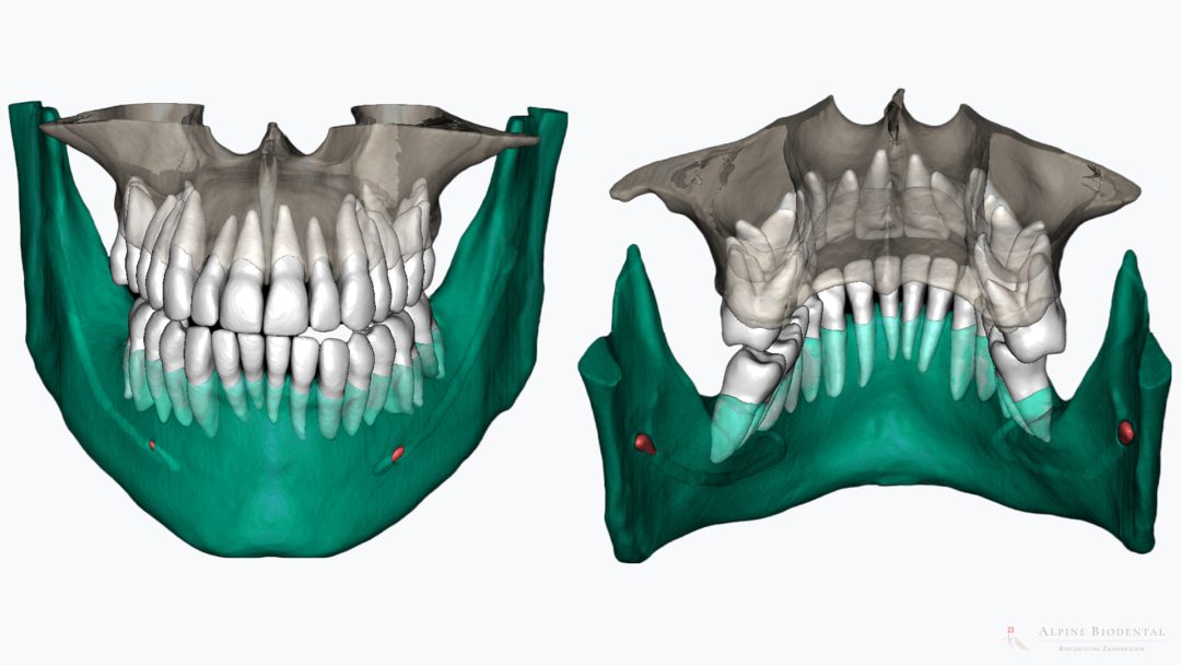
Inferior Alveolar Nerve
The inferior alveolar nerve is commonly at risk of damage during surgeries and implantations in the lower jaw. The nerve runs bilaterally in the lower jaw. Damage leads to sensory disturbances in the teeth, lower lip, and chin on the corresponding side. Arteries and veins always accompany the inferior alveolar nerve. In the images, the entry and exit points of the inferior alveolar nerve are highlighted in red.
Nerve in the Upper Incisors
This nerve can be damaged during implantations in the area of the middle upper incisors, causing sensory disturbances in the front palate. Similar to the lower jaw, nerves and blood vessels run together through the palate. The image depicts the course of this anatomical structure, with the exit point on the palate marked in red.
Procedure of Cone Beam Computed Tomography
CBCT creation is straightforward. After removing metal objects like earrings, necklaces, or dentures and wearing a lead apron, the 3D X-ray image is taken. The patient sits with support for the forehead and chin, keeping the head still for a few seconds. During this brief time, the X-ray tube and sensor move around the head, creating a three-dimensional image of anatomical structures. The CBCT can be accessed within seconds, allowing the dentist to diagnose alongside the patient.
Radiation Exposure in CBCT
The radiation exposure in CBCT depends on parameters, the volume size of the 3D X-ray, and the CBCT device used. Therefore, it’s crucial to set the parameters correctly and choose the ideal volume size based on the individual situation. The CBCT device should be of the latest generation and high quality, as technological advancements and device quality can minimize radiation dose. Generally, the radiation exposure of CBCT is approximately ten times lower than that of a head CT scan. We use the Planmeca ProMax Mid, which features the Planmeca Ultra Low Dose protocol, allowing the creation of high-quality 3D images with minimal radiation exposure (approximately 0.021 millisieverts or mSv). To put this in perspective, the average annual radiation dose for a person in Switzerland is about 5.6 mSv.
Dr. med. dent. Rebekka Hueber
A CBCT offers an accurate diagnosis before complex operations. Our team will be happy to assist you.

FAQ on the Topic: CBCT
The DVT 3D X-ray is an important diagnostic tool for complex treatment planning and prior to implantation.
No, the injection of contrast media is not necessary.
Yes, we can send you the complete 3D images or give them to you on a USB stick. You can view the three-dimensional X-ray at home using the enclosed viewing program.
No. The creation of CBCTs makes sense if the practice is oriented towards oral surgery.
Yes, all teeth (including the wisdom teeth) can be fully imaged in one scan.
No. CBCT is a completely painless examination.
Further information
Further information intended to give a better overview of the topic.